 |
MOUTH PART of a
male
Myrmarachne malayana spider 1- Palp/Palpus |
蜘蛛目錄
蜘蛛圖鑑
Spider Anatomy
|
|
MOUTH PART of a
male
Myrmarachne malayana spider 1- Palp/Palpus |
| 腹部 = abdominal, ventral, Ventral View 背部 = back or top, dorsal, Dorsal View 側視圖 = Lateral view |
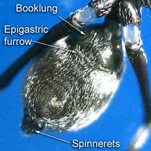 |
Myrmarachne malayana Male 6mm 2016-10-03 MON 15:21 SEMARAK ASIA
Booklung |
 |
Chrysso sp BOMBALAI Female ♀3mm 2015-09-05 FRI 12:17 BOMBALAI 1- Transparent white sternum (breast plate) 2- A pair of book lungs with white cover. 3- Abdomen oval, shorter than wide and armed with one line of 10 black seta at the tip edge. No obvious female epigynum. 4- Spinnerets yellowish as the ventral abdomen. |
 |
Gea
sp BUFFERZONE Male ♂4mm 12-8-2015 WED 17:48 BUFFER ZONE Ventral View Male pedipalp : 1- Bulb containing the coiled sperm duct. Prosoma (cephalothorax) - The first body part.
|
| Arachnids have two body parts as opposed to
insects with three. The first part is the prosoma and the second
part is the abdomen.
The prosoma, usually referred to as the cephalathorax, as it consists of a fused cephalic (head) and thorax which may be separated by a distinct or indistinct cervical groove and the fovea, a depression to which the stomach sucking muscles are attached. The shape of the fovea and cervical groove are important diagnostic features for the identification of spiders into their respective taxa. Chitin is responsible for this part of the exoskeleton being hard and inflexible. The prosoma houses various external appendages:
The opisthosoma contains the respiratory organs, heart, various spinning glands, the midgut, and ovaries for egg production.
|
|
Embolus |
1st row anterior eyes
Full Dorsal View
Body Dorsal View
Full Ventral View
Body Ventral View

Booklung
Epigastric furrow

Epigynum
Fang
Endite
Labium
Sternum (breastbone)
Booklung
Spinnerets
Palp
Booklung Cover
Prosoma (cephalothorax)
Opisthosoma (abdomen)
Male gonopore
Epigyne (epigynum) is the external genital structure of female spiders. As the epigyne varies greatly in form in different species, even in closely related ones, it often provides the most distinctive characteristic for recognizing species. It consists of a small, hardened portion of the exoskeleton located on the underside of the abdomen, in front of the epigastric furrow and between the epigastric plates.[1]
Maxillae (endites) or coxae of the pedipalps.
Calamistrum. The more or less extensive row of curved hairs on the hind
metatarsi, used to comb the silk from the cribellum.
Carapace. The hard dorsal covering of the cephalothorax of a
spider.
Cephalothorax or prosoma = A fused head and thorax. The
united head and thorax of Arachnida and Crustacea.
Chelicerac. The pincerlike first pair of appendages of the arachnids; in spiders
two-segmented, the distal portion or fang used to inject venom from enclosed
glands into the prey.
Chorion. The outer covering or shell of the spider or insect egg.
Coxa. The basal segment of the leg by means of which it is articu- lated to the
body.
Claw tufts. The pair of tufts of adhesive hairs present below the paired claws
at the tip of the tarsi of many spiders.
Colulus. The slender or pointed appendage immediately in front of the spinnerets
of some spiders; in other greatly reduced or seem- ingly missing; the homologue
of the anterior median spinnerets or cribellum.
Coxal glands. The excretory organs of arachnids, in spiders located opposite the
coxae of the first and third legs, that collect wastes into a saccule and
discharge them through tubes opening behind the coxae; homologous with the
nephndia of Peripatus, etc.
Cribellum. A sievelike, transverse plate, usually divided by a deli- cate keel
into two equal parts, located in front of the spinnerets of many spiders; the
modified anterior median spinnerets.
Cuticle. The hard outer covering of an arthropod.
Deutovum. The resting, spiderlike stage following the shedding of the chorion of
the egg; the second egg.
Dorsum. In general, the upper surface.
Ecdysis. The process of casting the skin; molting.
Endite. The plate borne by the coxa of the pedipalps of most spiders, used to
crush the prey; the maxilla.
Epigynum (female). The more or less complicated apparatus for storing the spermatozoa,
immediately in front of the opening of the internal reproductive organs of
female spiders.
Femur. The thigh; usually the stoutest segment of the spider's leg, articulated
to the body through the trochanter and coxa and bear- ing the patella and
remaining leg segments at its distal end.
Genitalia. All the genital structures.
Hackled band. The composite threads of the cribellate spiders, spun by cribellum
and combed by the calamistrum.
Carapace = Carapace is a protective cover over
the cephalothorax.
Chelicerae = The mouthparts of the of a spider. Chelicerae in spiders are hollow
and contain venom glands, and are used to inject venom into prey.
Abbreviations :
AL= abdominal length, ALE= anterior lateral eye, AME=
anterior median eye, AW= abdominal width, CL= cephalothoracic length, CW=
cephalothoracic width, PLE= posterior lateral eye, PME= posterior median eye,
TL= total length.
|
|
Common Spiders of Borneo |
