Last Updated on Sunday, 17 September, 2023 03:14:12 PM
INDEX >
Dragonflies of Borneo >
The abdomen of odonates
The abdomen of odonates
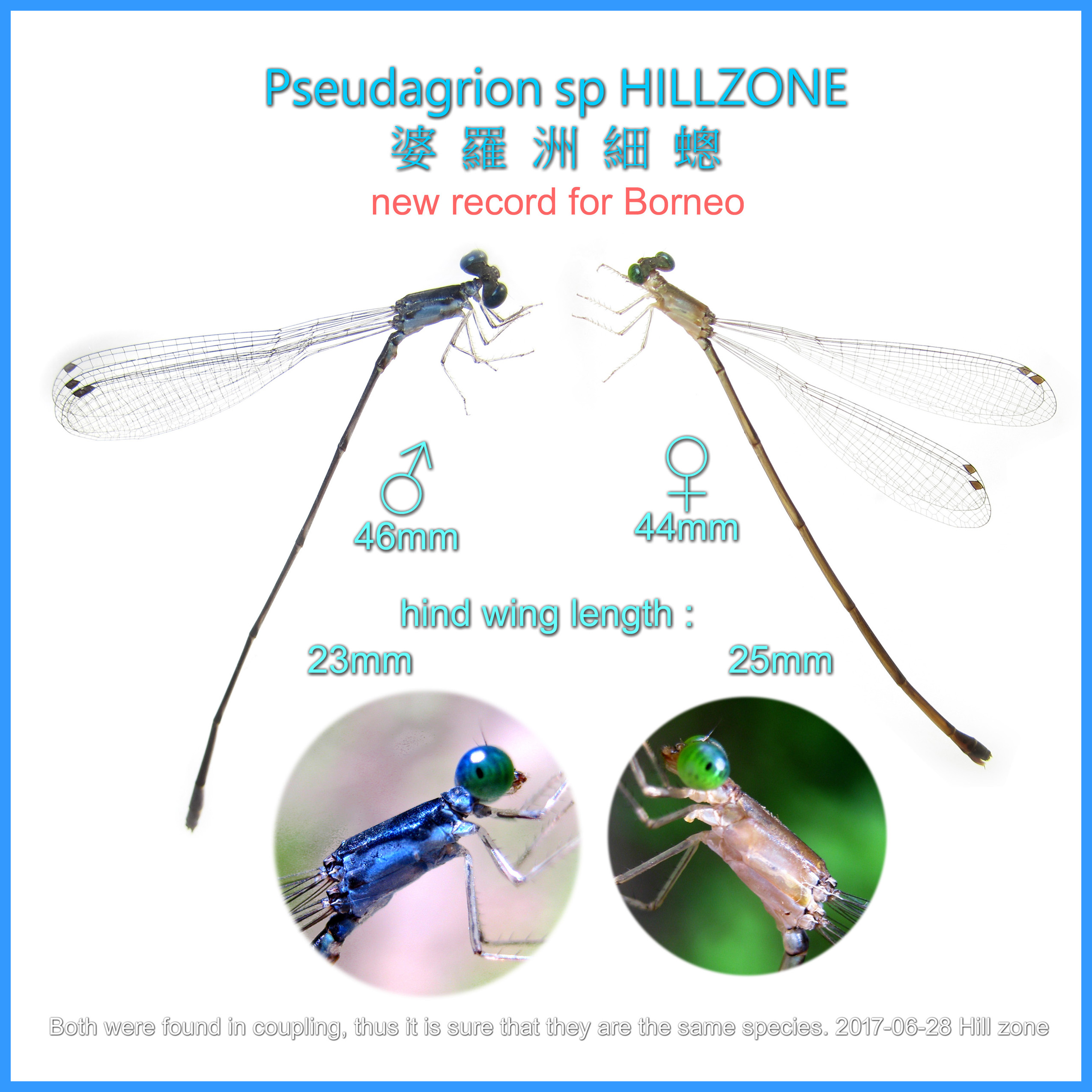
ABDOMEN DIFFERENT BETWEEN MALE AND FEMALE DRAGONFLIES
Most male dragonfly abdomen is often narrower at segment 3, whereas the female
abdomen is more robust.
1- Male abdomen is often narrower at segment 3
2- Female abdomen is more robust
For the female dragonflies , genitalia are located underneath the abdomen
between segment 8 and 9.
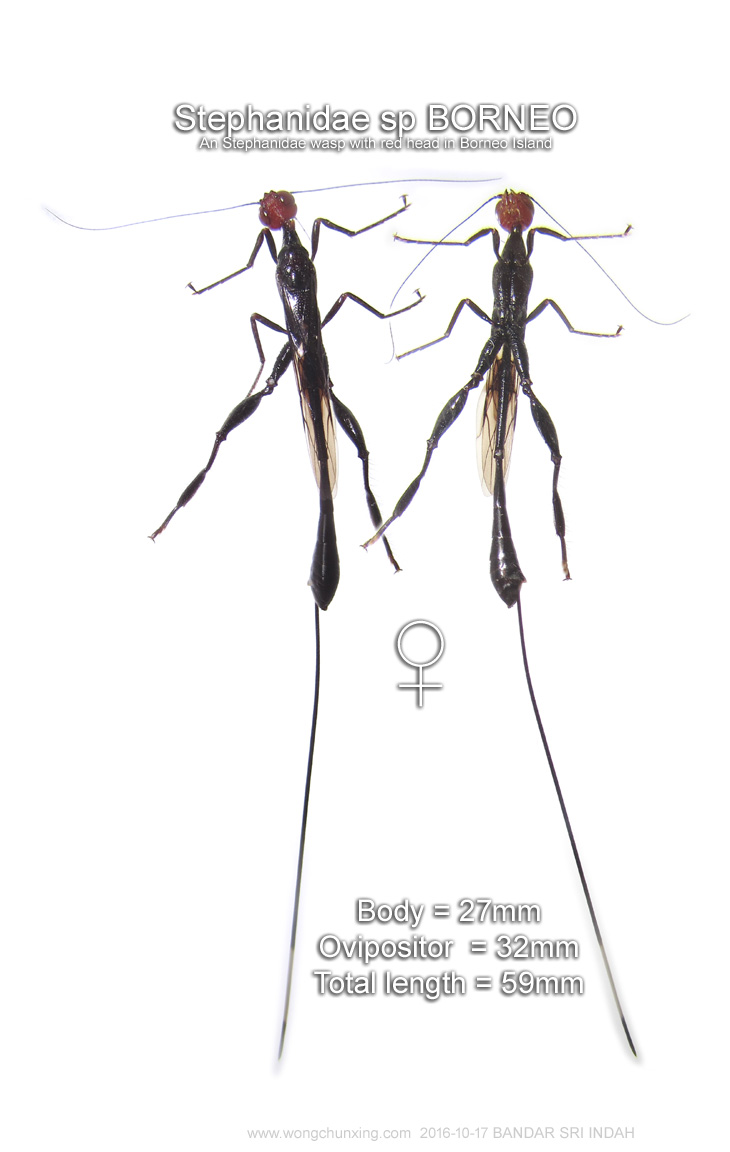
Zygoptera and some Anisoptera (Aeshnidae and Cordulegaster) have an ovipositor,
surrounded by valvae.
An ovipositor is used to stick eggs in plants, wood or mud. These species are
called endofytic.
Dragonfly Species that don’t have an ovipositor (exofytic) simply drop their
eggs into the water.
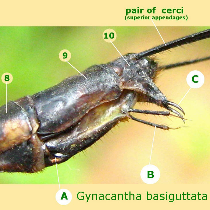
At the back of segment 10, at the tip of the abdomen, the appendages are
located. These are especially clearly visible at male odonates.
Male Zygoptera have two pairs of anal appendages: the upper appendages
(appendices superiors) and the lower appendages (appendices inferiors).
Anisoptera males only have upper appendages, plus one single small knob that is
also called lower appendage.
Female odonates only have very small appendages, which are functionless. These
are called cerci.
The appendages of male odonates have an important function during the
copulation: with these appendages, the males hold the females behind the eyes (Anisoptera)
or at the back edge of the prothorax (Zygoptera). The shape of the appendages of
a species is typical for that species and corresponds with the shape of the
female’s prothorax (Zygoptera), which prevents bastardizing. For Anisoptera, the
hamulus prevents bastardizing: only female’s genitalia of the same species fit.
Very rarely however, bastardizing occurs.
Abdomen
The abdomen is divided into 10 segments, numbered from 1 at the base to 10 at
the tip. The major features of the abdomen useful for identification are the
sexual appendages, extending from the terminal segments, and the secondary
genitalia of males.
Secondary genitalia of males are located under abdominal segment 2. Of the many
intricate structures that make up the secondary genitalia, the most useful to
learn for field identification are the hamules, which are hook like structures
on most dragonflies; they are visible with a hand lens and are often species
specific in shape.
At the primary genitalia of males are the terminal appendages of males, which
are used to grasp females by the rear of the head (in typical dragonflies) or
the thorax (in damselflies) during mating, are often important features for
distinguishing look-alike species.
It is therefore worthwhile to learn the names and positions of these structures.
The cerci (singular, cercus) are the pair of upper (superior or dorsal)
appendages. When the male curls his abdomen down and forward to grasp the
female, the cerci curl under the upper rim of the head (in typical dragonflies)
or contact the mesostigmal plates (in damselflies).
Male cerci are often relatively large appendages with hooks or spines for
grasping. The cerci of females are typically simple, cone-shaped or leaflike
structures and only occasionally of use in field identification.
The lower (inferior or ventral) grasping appendages of male damselflies and
typical dragonflies are different in nature. Male damselflies have a pair of
structures, the paraprocts, that grasp the prothorax during tandem linkage. In
some species, these are relatively large and strongly hooked. In nearly all
other odonates, the paraprocts are relatively inconspicuous, small, rounded
structures. The inferior grasping appendage of typical male dragonflies is a
single epiproct (strongly forked in some species) that grasps the top of the
head or eyes of females. This structure typically is not a conspicuous feature
on female dragonflies or most damselflies. However, on male dancers (Argia), the
shape and size of the epiproct—which appears as a small lobe projecting rearward
from the upper middle rim of segment 10—relative to the shape of the projecting
pads (called tori [singular, torus]) on either side of it are of use in
identifying some species.

The females of all damselflies and some dragonflies (darner and petaltail families
[Aeshnidae and Petaluridae]) have a fully formed ovipositor, which is a
complicated structure containing paired valves and cutting blades, on the
underside of abdominal segments 8 and 9.
The ovipositor is used to insert eggs into plant tissue,mud, or other substrate.
Some species have a stylus, which is a thin, needlelike projection, at the end
of each of the two valves of the ovipositor. Species without a true ovipositor
(most of the typical dragonflies in our area) have a more or less well developed
vulvar lamina, a plate that extends rearward from segment 8 to cover part of the
undersurface of segment 9. This plate, which is often distinctly bilobed, may be
used to carry egg masses or otherwise aid in the dispersal of eggs. In the
spiketail family (Cordulegastridae), the vulvar lamina is highly modified to form
a spikelike structure that inserts eggs, much as a true ovipositor might, into
aquatic substrates.
Some species of damselflies have a vulvar spine on the rear lower margin of
segment 8 that projects over the genital opening at the base of segment 9. You
will occasionally find odonates, especially damselflies, with tiny red “beads”
attached, often in small clusters, to the undersurface of the thorax or abdomen.
These are not part of the odonate but rather are larvae of parasitic water
mites, which hitch a ride on odonate larvae and then make the transfer to the
adult form at the time of emergence. The mite larvae attach themselves to the
hardening body, sucking fluids from their host. When the adult odonate later
comes into contact with water— for example, during oviposition—the mites detach
and return to the water to complete their life cycle.
There are few other insects that might be mistaken for odonates. Perhaps most
likely to cause confusion are adults of the antlion family (Myrmeleontidae,
order Neuroptera), which resemble adult damselflies but have noticeably longer,
clubtipped antennae.
The abdomen always has ten segments. Segments 1 and 2 appear to be integrated
into the thorax and are sometimes difficult to tell from the thorax. To find a
particular segment, it is usually best to start with segment 10, far out at the
tip, and count backwards. Because of its segmented nature, the abdomen is very
flexible and is able to arch up or down (but not side to side).
The female terminal appendages consist of a pair of cerci, which have little or
no function. In some species, namely the Shadow Darner, they are very brittle
and tend to break off. Underneath segment 8 there is either an ovipositor or a
subgenital plate, depending upon the species. Both structures are for laying
eggs and extend over segment 9 and possibly beyond.
The male abdomen is often narrower (“waisted”) at segment 3, whereas the female
abdomen is almost always more robust.
The abdomen of odonates, which consists of 10 segments plus the rudiment of
an 11th segment, is remarkably long and slim, rarely very broad and short. The
abdomen houses the internal organs and the genitalia. It also acts as a steer
and helps keeping balance in flight.
Segment 1 is very short and not visible from above. Segment 3 to 7 are quite
long, the others are shorter. Segment 10 carries a few projections called anal
appendages.
Each segment consists of an upper side or tergite and a bottom side or sternite,
connected by pleurites at the flanks. The tergites are much bigger and harder
than the sternites, which are quite weak and flexible.
The genitalia of odonates are very complex. Males have their sperm production
organ in the 9th segment (like any other insect) but their copulation organ
(that acts as a penis) is located underneath segment 2. These organs are not
interconnected, so the male has to transfer his sperm from the producing organ
to the penis before the actual copulation takes place!
The penis (called ligula) is an important characteristic for the recognition of
male Zygoptera in case of doubt.
The hamulus
- A pair of hooks in segment 2
For libellulidae (a group of Anisoptera), the hamulus can be used for species
recognition. The hamulus is a set of hooks situated at the sternite of segment
2, used to hold the female’s genitalia during copulation.
Prior to the selection of a willing female, the male will transfer sperm from
his testes located on the underside of abdominal segment 9 to his hamulus
located on the underside of segments 2 and 3. This is accomplished by simply
arching the abdomen until the undersides of the appropriate segments make
contact.
Mating is normally initiated by the male who, with the grace of a professional
wrestler, uses his legs to grasp the female by her head and thorax. Curving his
abdomen forward, he uses his two cerci and the lower epiproct as a clamp and
clasps the female by the back of the head. They are now “in tandem.” Mating is
accomplished by the male arching his abdomen downward while the female arches
her abdomen toward the male’s hamulus. Once connected, the pair is in the wheel
position or “in copula.” Still connected, the pair will usually fly up to the
safety of treetops to mate, although some species do copulate in mid flight. The
male commences a purging of the female’s genital opening. He uses the hamulus to
remove, squash or push out of the way any sperm that the female may still be
carrying from prior matings with other males. This process ensures his genetic
investment in the clutch of eggs that the female will soon lay. The time needed
to complete fertilization ranges from 15 seconds to well over an hour.
The male testes are located in segment 9. Due to the unique nature of dragonfly
copulation, the male must transfer sperm to his secondary genitalia, called the
hamulus, located in the underside of the second and third segments.
The hamulus is like a set of “surgical tools” that a male uses for removing
sperm left by other males during previous matings.
Other parts of the hamulus are then used by the male to fertilize the female
with his own sperm. The terminal abdominal appendages of the male are called
claspers. The claspers are formed by a pair of upper appendages, called cerci,
and a single lower appendage, an epiproct. In some species, the males possess
auricles on the sides of segment 2 whose function is to help direct the female’s
genitalia to a proper fit with the male’s secondary genitalia during copulation.
The 3 families of dragonflies found in Borneo Island:
1 Family of Aeshnidae

CHART OF DRAGONFLIES OF BORNEO
RELATED TOPICS
INTRODUCTION TO THE DAMSELFLIES OF SABAH, BORNEO ISLAND
Insects are diverse and dominant inhabitants of the tropical rainforests in Borneo Island. New species are discovered too often. Entomologists are still struggling to cope with the documentation of tropical insect diversity.
Most of the common dragonflies in Sabah are red coloured, especially from the family Libellulidae. Some red pecies are even confused as same species, for example the three species of Genus Neurothemis.

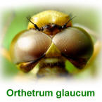
The compound eyes of dragonflies
Dragonflies and damselflies have large compound eyes that can see in all directions. When the compound eye is magnified several hundred times, each individual facet (ommatidium) is shown to be hexagonal in shape.
Ovipositor (Vulvar Lamina)
of Female Dragonflies and Damselflies
Female dragonflies have either one of the two method of depositing eggs from
the abdomen:
1- using Ovipositor Structure
2- using Vulvar Lamina
Male do not have an ovipositor. Instead male dragonfly and damselfly have appendages.
|
GO FURTHER FLORA Borneo has 150 species of wild fig trees. Most of them are found in forests of Sabah. FISHERIES Prawn farming is a main commercial activities in Tawau. Spawners from Tawau are graded the best in Malaysia. For decades, Tawau has been exporting high-grade tiger prawns to several countries such as Korea, Japan, Taiwan, China, Vietnam, Singapore, Egypt and Australia. |
|
|
Damselflies of Borneo |